Author: Julie
-
Waverley House Accelerator for Mental Illness and Addiction Research – Now Recruiting Five Early-Career Mental Health Research Leaders
The University of Ottawa Institute of Mental Health Research (IMHR) and The Royal are pleased to launch the Waverley House Accelerator for Mental Illness and Addiction Research — a transformative initiative designed to recruit and support the next generation of research leaders in mental illness and addiction science. Supported through a landmark investment from the
-

CAN 2026 Election results
We are happy to announce the results of the 2026 elections – thanks to all the members who participated! Elected member begin their mandate on June 1, 2026 Elected Board members Jo Anne Stratton Vice-President-Elect Secretary-Elect
-

Congratulations to the winners of the 2025 CAN- CIHR-INMHA Brain Star Awards!
The Canadian Association for Neuroscience (CAN) and the Canadian Institutes of Health’s Institute of Neurosciences, Mental Health and Addiction (CIHR-INMHA) are proud to announce the winners of the 2025 Brain Star Awards. The CIHR-INMHA Brain Star awards, administered by the Canadian Association for Neuroscience, are awarded to students and trainees who have published high impact
-
Postdoctoral Position – Neuroscience/Neuropharmacology – 2 years – Manitoba
St Boniface Hospital Albrechtsen Research Centre& University of Manitoba POSTDOCTORAL POSITION Neuroscience/Neuropharmacology – 2 years Project: Using Sonogenetics to Replace Pharmaceuticals A US government funded project will mobilize ultrasound to activate genetically modified ion channels to drive nerve repair and suppress pain. Neuronal cell lines and adult primary sensory neurons will be transduced with AAVs
-
PhD student position in neuroscience of chronic pain – University of Guelph
Are you interested in exploring brain circuits and gene expression networks? Do you want to determine the roles astrocytes play in neuronal plasticity? The Descalzi lab is looking for a PhD student to join their team at the University of Guelph! Job Title: PhD Student Position Location: University of Guelph, Guelph, Ontario, onsite About
-
Postdoc position in neuroscience of chronic pain – Guelph University
Are you interested in exploring brain circuits and gene expression networks? Do you want to determine the roles astrocytes play in neuronal plasticity? The Descalzi lab is looking for a postdoctoral researcher to join their team at the University of Guelph! Job Title: Postdoctoral Fellow Position Location: University of Guelph, Guelph, Ontario, onsite About
-
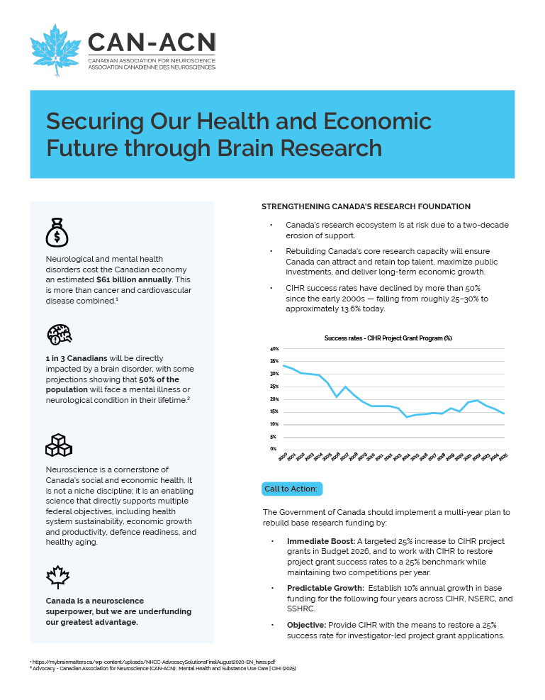
Read CAN’s submission to pre-budget consultations
The House of Commons Standing Committee on Finance is inviting Canadians to participate in its annual pre-budget consultations process. A report on these consultations will be tabled in the House of Commons. The committee will accept written briefs through the committee’s website until Friday, May 22, 2026, at 11:59 p.m. Eastern Standard Time. CAN submitted
-
Postdoctoral Fellow / Senior Data Scientist / Biostatistician – Memorial University of Newfoundland
Position Title: Postdoctoral Fellow / Senior Data Scientist / BiostatisticianNeuroimmunology & Multiple Sclerosis ResearchMemorial University of Newfoundland – Faculty of Medicine Location: St. John’s, Newfoundland and Labrador, Canada (remote or hybrid work arrangements may be considered)Duration: 1 year (renewable, contingent on performance and funding)Start Date: Spring-Summer 2026 The Translational Neuroimmunology Laboratory invites applications for a highly motivated to join
-
Assistant or Associate Professor, Department of Human Anatomy & Cell Science
Posting Title: Advertisement For Assistant or Associate Professor Position number: 37564 Type: Assistant or Associate Professor Department: Human Anatomy & Cell Science Faculty: Rady Faculty of Health Sciences Posting start date: May 4, 2026 Posting close date: June 5, 2026 For more information please contact: Jacki Armstrong jacki.armstrong@umanitoba.ca Position description: The Department Human Anatomy &
-
CAN Parliament Hill Day 2026
The Canadian Association for Neuroscience held its 2026 Parliament Hill Day on April 14, 2026. It was an opportunity for Canadian neuroscientists to highlight the important work they do for Canada and the importance of government support for Canadian Research. The meetings In total, CAN had 19 meetings, including CAN participants We are grateful to