Category: Awards
Awards advertisements
-
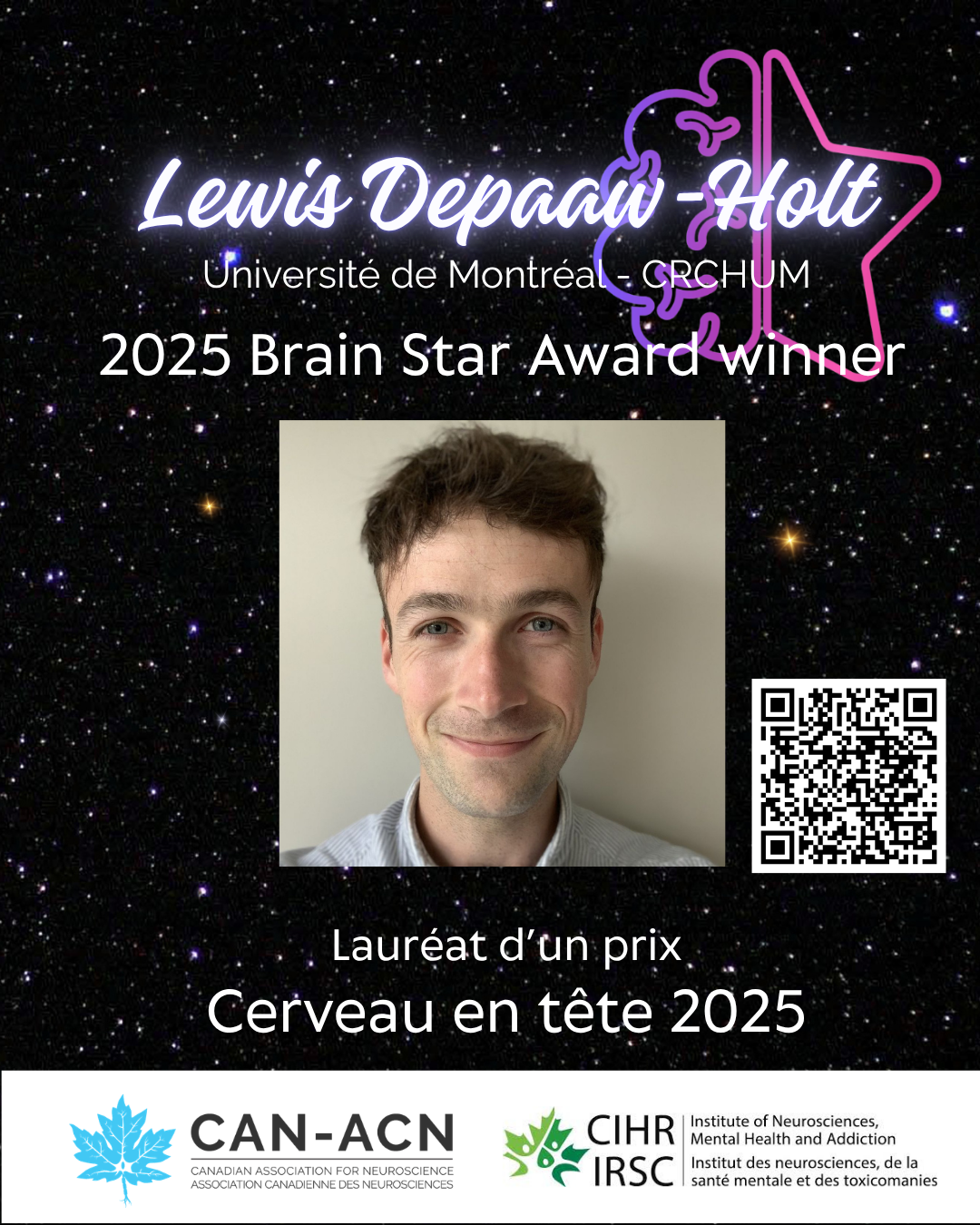
Brain Star Award winner feature: Lewis Depaaw-Holt, Université de Montréal
Astrocytes play key roles in sex-specific changes in brain and behaviour caused by early-life stress In this study, Lewis Depaauw-Holt, working in the laboratory of Dr. Ciaran Murphy-Royal at Université de Montréal, investigated how early life stress influences brain and behaviour. They specifically aimed to elucidate the role of a non-neuronal brain cell, the astrocyte.
-

Brain Star Award feature: Christopher Daniel Morrone – Centre for Addiction and Mental Health, University of Toronto
Brain cleaning deficiencies are linked to sleep loss at the early stages of Alzheimer’s disease Autophagy involves the recycling of the cell’s own components, such as damaged proteins, and is a highly regulated mechanism that is essential for maintaining cellular health. Research done by Christopher Daniel Morrone working with Dr. Wai Haung Yu at the
-
Congratulations to the winners of the 2025 CAN- CIHR-INMHA Brain Star Awards!
The Canadian Association for Neuroscience (CAN) and the Canadian Institutes of Health’s Institute of Neurosciences, Mental Health and Addiction (CIHR-INMHA) are proud to announce the winners of the 2025 Brain Star Awards. The CIHR-INMHA Brain Star awards, administered by the Canadian Association for Neuroscience, are awarded to students and trainees who have published high impact
-
Brain Star Award feature: Sergio Crespo-Garcia, Maisonneuve-Rosemont Hospital Research Center
As the last Brain Star Award Feature of this series, we are proud to present the first place winner of the 2024 competition, Dr. Sergio Crespo-Garcia. He is also the winner of the Marlene-Reimer Award for 2024. Congratulations! Discovery of a new therapeutic avenue to protect vision in diabetic patients. Diabetes is a silent epidemic
-
Brain Star Award Feature: Christina You Chien Chou – McGill University
An optomapping approach to better understand connections in the visual cortex of the brain In the brain, information is passed from neuron to neuron via connections called synapses. Synaptic dysfunction unsurprisingly underlies many neurological diseases, such as autism, schizophrenia, and epilepsy. Understanding how synapses are wired up in a cell-type-specific way is fundamental to understanding
-
Brain Star Award Feature: Justine Hansen, McGill University
Studying how the deepest regions of the brain contribute to brain activity The brainstem is a structure which is crucial for survival and consciousness, yet it is typically excluded from live human brain mapping efforts due to the difficulties in recording and analysing activity in this small region which sits deep at the base of
-
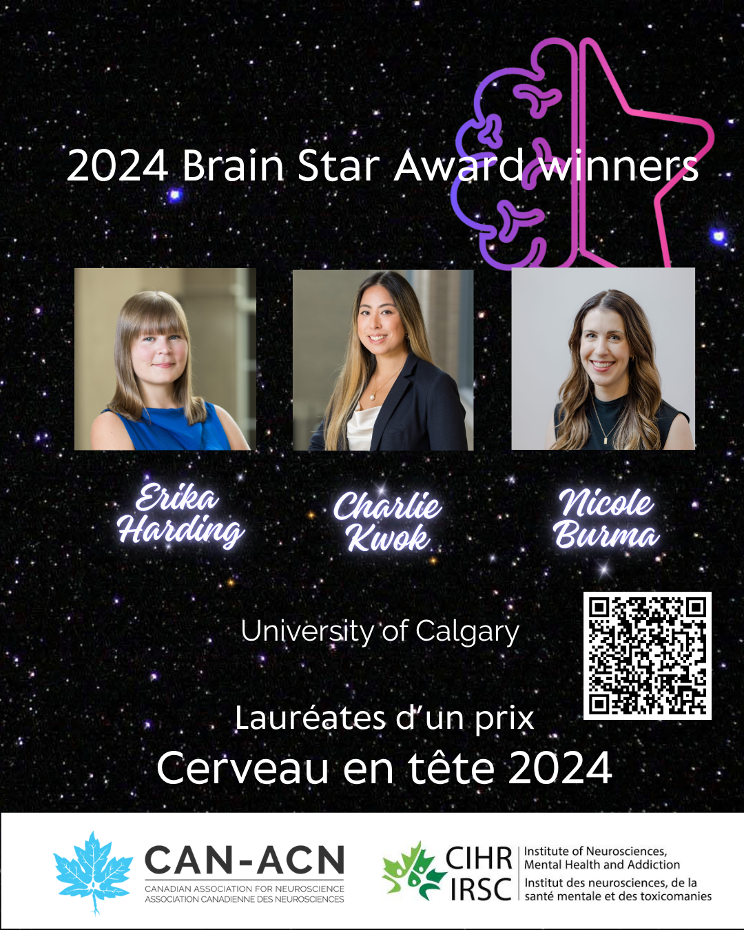
Brain Star Award feature: Erika Harding, Nicole Burma, Charlie Hong Ting Kwok, University of Calgary
Identification of the Pannexin-1 channel in the brain as a target to treat opioid withdrawal symptoms Opioids remain one of the most effective analgesics, with 10-15% of Canadians receiving opioid prescriptions per year. However, opioids are also highly associated with substance use disorders and overdose related deaths. Last year alone, over 7000 Canadians passed away
-
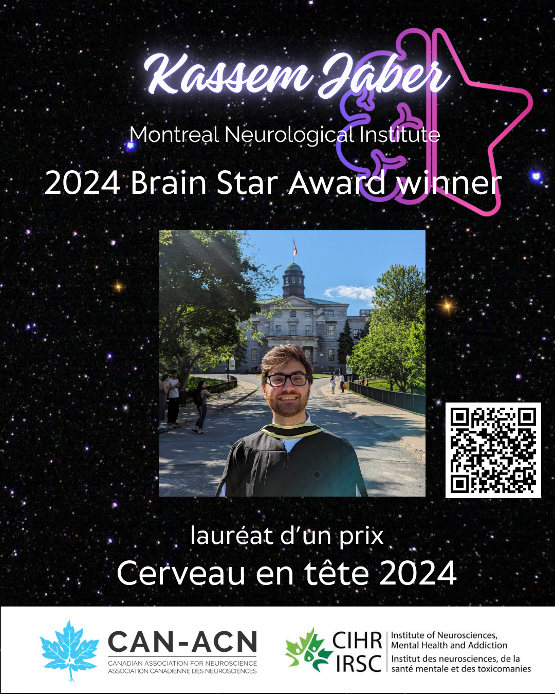
Brain Star Award Feature: Kassem Jaber, Montreal Neurological Institute
A new framework to assess placement of electrodes in the brain for epilepsy surgery Epilepsy is a chronic condition that is characterized by spontaneous recurring seizures. In clinical practice, the region which generates seizures is called the epileptic focus. The location of the focus can be localized by electrical measurement of brain activity, known as
-
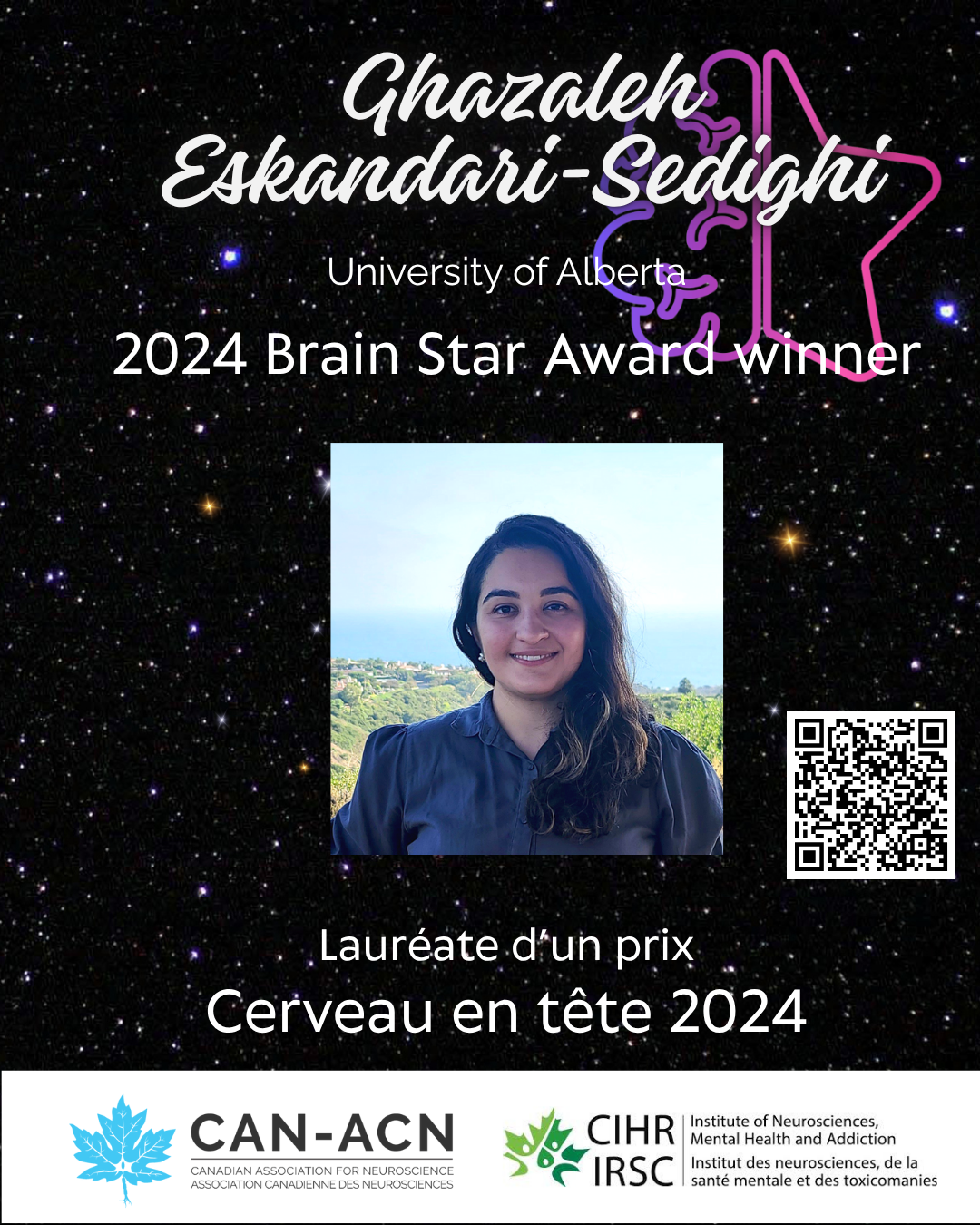
Brain Star Award Feature: Ghazaleh Eskandari-Sedighi, University of Alberta
Identification of CD33m as a new protective factor in Alzheimer’s Disease development. Immune cells in the brain, called microglia, are thought to be critical in Alzheimer’s disease (AD) development through numerous functions, including their ability to remove amyloid beta (Aβ), which is protein that accumulates in the brains of AD patients. In this study, Ghazaleh
-
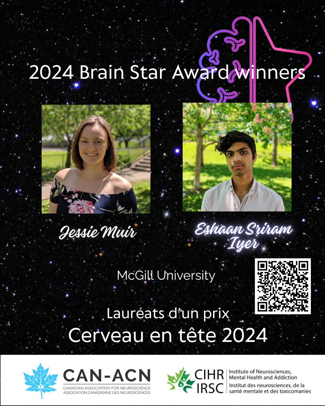
Brain Star Award Feature: Jessie Muir and Eshaan Sriram Iyer, McGill University
Discovery of differences in encoding threat discrimination in the brain of males and females Learning to predict threat is essential, but equally important—yet often overlooked—is learning about the absence of threat. This study by Drs. Jessie Muir and Eshaan Sriram Iyer, working in the laboratory of Dr. Rosemary Bagot at McGill University, looks at mechanisms