Category: News
-
Brain Star Award winner feature: Lewis Depaaw-Holt, Université de Montréal
Astrocytes play key roles in sex-specific changes in brain and behaviour caused by early-life stress In this study, Lewis Depaauw-Holt, working in the laboratory of Dr. Ciaran Murphy-Royal at Université de Montréal, investigated how early life stress influences brain and behaviour. They specifically aimed to elucidate the role of a non-neuronal brain cell, the astrocyte.
-

Brain Star Award feature: Christopher Daniel Morrone – Centre for Addiction and Mental Health, University of Toronto
Brain cleaning deficiencies are linked to sleep loss at the early stages of Alzheimer’s disease Autophagy involves the recycling of the cell’s own components, such as damaged proteins, and is a highly regulated mechanism that is essential for maintaining cellular health. Research done by Christopher Daniel Morrone working with Dr. Wai Haung Yu at the
-
Read CAN Connection – Summer 2026
We invite you to read our June newsletter!
-
CAN 2026 Election results
We are happy to announce the results of the 2026 elections – thanks to all the members who participated! Elected member begin their mandate on June 1, 2026 Elected Board members Jo Anne Stratton Vice-President-Elect Secretary-Elect
-
Congratulations to the winners of the 2025 CAN- CIHR-INMHA Brain Star Awards!
The Canadian Association for Neuroscience (CAN) and the Canadian Institutes of Health’s Institute of Neurosciences, Mental Health and Addiction (CIHR-INMHA) are proud to announce the winners of the 2025 Brain Star Awards. The CIHR-INMHA Brain Star awards, administered by the Canadian Association for Neuroscience, are awarded to students and trainees who have published high impact
-
Maria Ioannou wins the 2026 CAN New Investigator Award for groundbreaking research on lipid metabolism in the brain
The Canadian Association for Neuroscience is very proud to announce Dr. Maria Ioannou, from the University of Alberta, has been awarded the CAN 2026 New Investigator Award for her innovative work that has revealed new mechanisms by which neurons expel, transfer, and detoxify peroxidized lipids, and how glia act as indispensable metabolic buffers that preserve
-
Read the latest CAN Connection Newsletter
We invite you to read the latest CAN Connection Newsletter – March 2026 Message from the President, Douglas Zochodne CAN Board of Directors – Call for nominations now open Advocacy updates CAN Meeting updates CAN Satellites and partner sessions Opportunities and Resources
-
Read our letter to Prime Minister Mark Carney
CAN President Douglas Zochodne sent a letter on behalf of CAN to Prime Minister Mark Carney on Neuroscience research and CIHR Project Grant Operations shortfalls. Read the letter here:
-
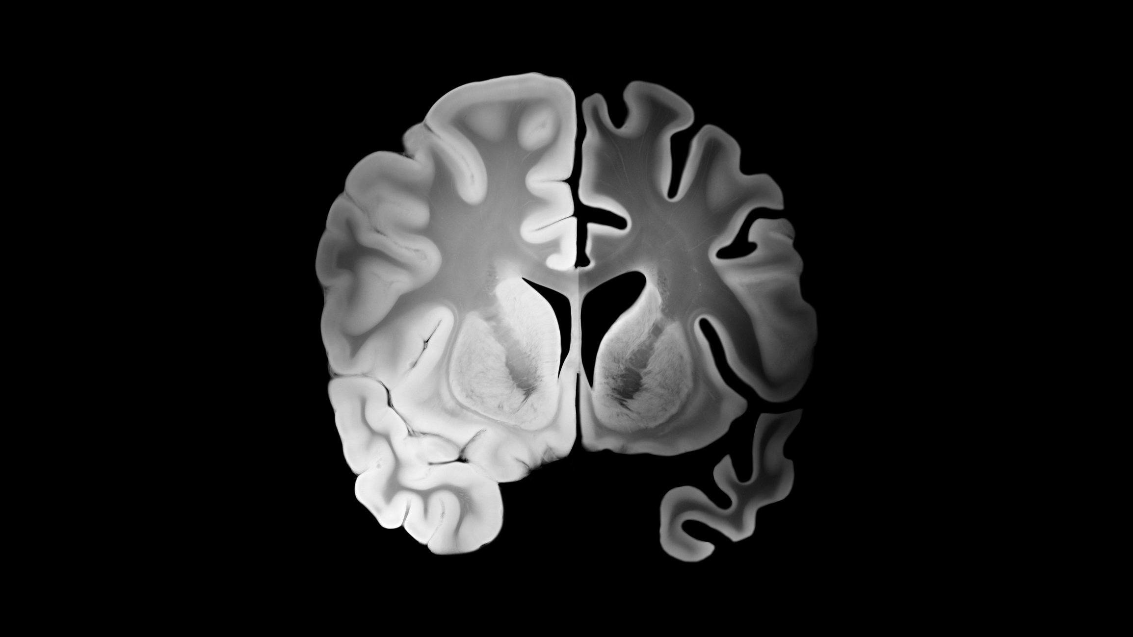
University of Toronto researchers identify potential biomarker linked to MS progression
Study co-leaders are Jen Gommerman, a professor and chair of immunology at the U of T’s Temerty Faculty of Medicine, and Valeria Ramaglia, a scientist at the UHN’s Krembil Brain Institute and a U of T assistant professor of immunology. Story by Betty Zou, UofT News Researchers at the University Health Network and University of
-
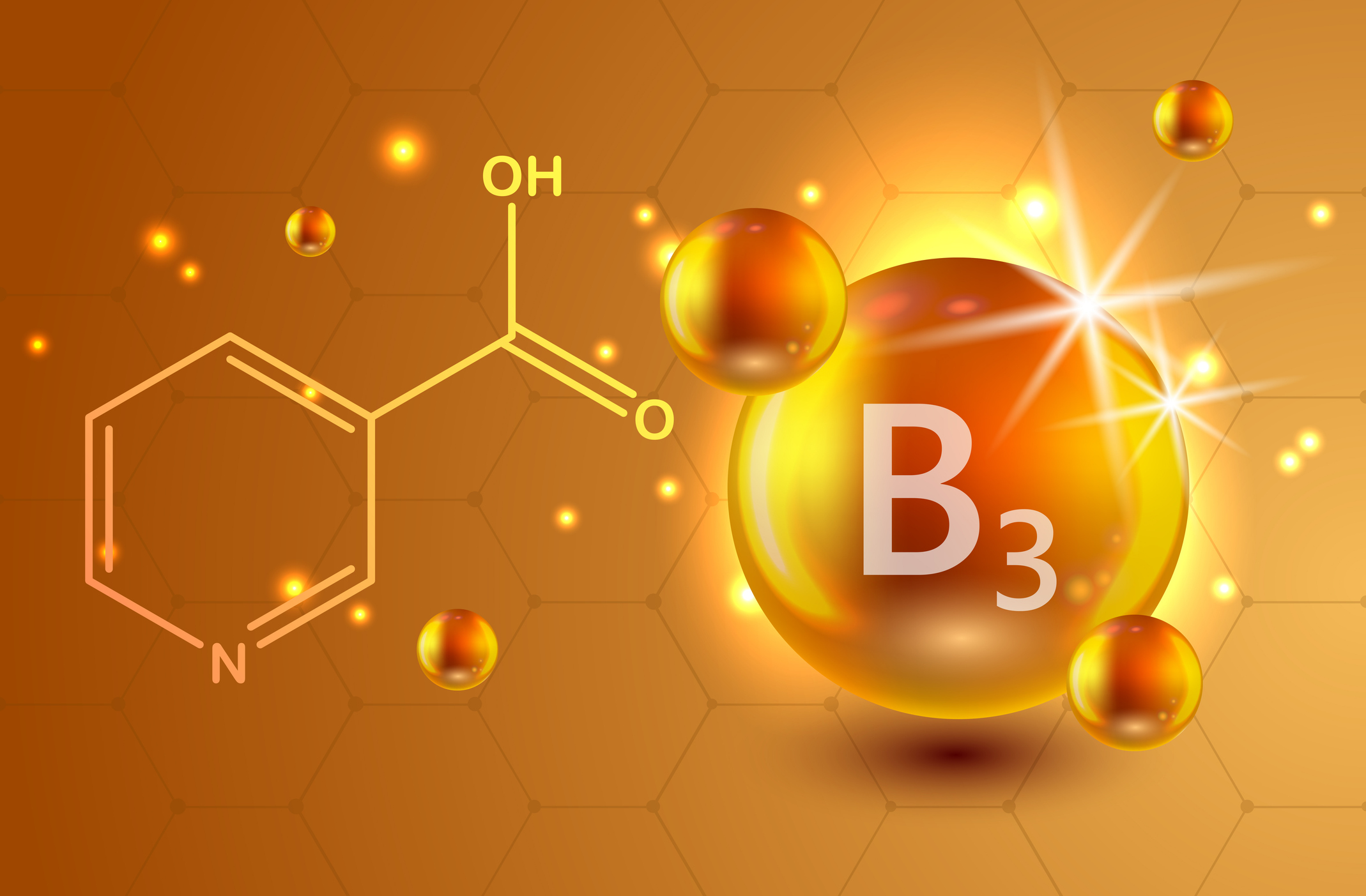
UCalgary study investigates the use of common vitamin to treat the aggressive brain cancer glioblastoma
Findings indicate vitamin B3 looks promising to help rearm a compromised immune system Story by Kelly Johnston, Cumming School of Medicine, University of Calgary published on Feb 11, 2026 University of Calgary and Hotchkiss Brain Institute researchers Voon Wee Yong and Gloria Roldan Urgoiti recently published a study in the Journal of Neuro-Oncology investigating whether adding